|
|||||||||||||||||||||||
|
Cardiac
Biomarkers 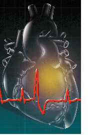 Every
year nearly 1.5 million individuals in the US suffer a heart attack (an
acute myocardial infarction, AMI), and of these approximately 500,000 die.
Ischemic heart disease is the principal cause of death in the US and involves
the consumption of enormous economic resources.1
Every
year nearly 1.5 million individuals in the US suffer a heart attack (an
acute myocardial infarction, AMI), and of these approximately 500,000 die.
Ischemic heart disease is the principal cause of death in the US and involves
the consumption of enormous economic resources.1
The most difficult challenge to emergency room physicians is the accurate triage (risk stratification) of the patients who make the 8 million visits to the emergency room for chest pain each year. Even though approximately 60 percent of those presenting with chest pain are admitted, a significant number (around 5 percent) of AMI patients go undetected and are released, a situation constituting the single largest cause of medical malpractice suits. Nevertheless, nearly one-half of admissions to the coronary care unit (CCU) are inappropriate. This unnecessary use of hospital resources contributes significantly to the billions of dollars spent on care for patients presenting with chest pain.1‚4 Cardiac markers provide information that is essential for accurate and timely triaging and diagnosis of AMI and for early, noninvasive detection of reperfusion following thrombolytic treatment. Pathophysiology
of AMI Pharmacological intervention with intravenous thrombolytic therapy to reopen occluded blood vessels has become a widely accepted treatment regimen that may avoid invasive procedures (angioplasty) among patients who present within the first 6 hours after infarction.1,3 Myocardial markers can be used to monitor the success of reperfusion noninvasively, as an alternative to coronary angiography. Although the latter has been considered to be the most definitive technique, it requires cardiac catheterization. The objectives in the emergency department are therefore to accurately triage patients presenting with chest pain as rapidly as possible to ensure that all and only the true AMI patients are admitted to the CCU and that appropriate treatment regimens are initiated in the shortest time possible after the infarct. The diagnostic procedures must have adequate sensitivity to maximize the probability of identifying the patients who have actually suffered an AMI (minimizing the number of false negatives), and adequate specificity to minimize unnecessary CCU admissions and their associated costs. Diagnostic
Tests This sensitivity limitation necessitates reliance on biochemical serum cardiac markers: proteins that leak into the circulation from damaged myocardial cells and serve as essential tools for triaging patients with indeterminate ECGs. Furthermore, cardiac markers also serve as powerful tools for the noninvasive assessment of myocardial reperfusion following thrombolytic therapy. As a result of reperfusion, trapped markers are washed out of the affected area and released into the circulation. A rapid rise of the markers during the 60- to 90-minute period after therapy signals successful reperfusion. Absence of a characteristic rise alerts the clinician to consider alternative therapies.1,3,4,6 Biochemistry,
clinical utility and limitations However, elevated levels may also be related to various skeletal muscle traumas and renal failure, and are therefore not specific for cardiac muscle injury. Serial determinations improve the specificity and predictive value of myoglobin for cardiac muscle injury. Sequential negative results rule out AMI, whereas 1- to 2-hour doubling times provide strong evidence for AMI. Despite the lack of cardiac specificity, myoglobin appears to best fit the role of an early marker for AMI. As is the case for the other cardiac markers, myoglobin also serves as a useful marker for the detection of successful reperfusion following thrombolytic therapy. Furthermore, the rapid clearance of myoglobin from the circulation (T1/2 is approximately equal to 3 hr) enhances its utility as an early marker for reinfarction. Creatine
kinase MB isoenzyme (CK-MB)1‚4,6,8,9
Although CK-MB has been the gold standard for detecting myocardial necrosis, it does have significant limitations and is not an ideal marker. The time to appearance of elevated levels is slower than that for myoglobin; CK-MB therefore does not meet the criteria for an early marker. Although CK-MB is < 1% of total CK in skeletal muscle, regenerating skeletal muscle following injury has a CK isoenzyme distribution that is similar to heart muscle composition, potentially leading to false-positive diagnosis of AMI. This lack of cardiac specificity can confound interpretation of CK-MB results when unrelated muscle trauma and disease processes are present in a patient being evaluated for AMI.9,10 As with myoglobin, serial determinations of CK-MB enhance its efficiency for the diagnosis of AMI and for assessing reperfusion following thrombolytic therapy. An additional limitation is related to the possible return of CK-MB to normal circulating levels within 48 hours after infarction. This has necessitated reliance on other markers, e.g., lactate dehydrogenase (LD) and the ratio of LD isozymes 1 and 2 (LD1/LD2), to evaluate patients arriving in the emergency department 48 hours after onset of chest pain.1-4,6,8,9 Cardiac
troponin I (cTnI)1‚4,6,8,10
The most important characteristic of cTnI, however, is its apparent absolute cardiac specificity. In contrast to all other known cardiac markers including cTnT, cTnI is not expressed in fetal, diseased or regenerating skeletal muscle.4,11 cTnI is not increased in patients with skeletal muscle or renal disease, and the absolute cardiac specificity of this marker allows its use for the diagnosis of perioperative myocardial infarction. Furthermore, this extraordinary cardiac specificity of cTnI has resolved risk stratification difficulties when interpretation of results for other cardiac markers was severely confounded by conditions unrelated to AMI.12 Among patients with acute coronary syndromes, cTnI levels have been reported to provide prognostic information useful for the early identification of patients with an increased risk of unstable angina progressing to AMI and death.5,13 Table
1.
cTnI
as the next gold standard?2,5,14 References 1. Keffer JH. Myocardial markers of injury. Am J Clin Pathol 1996;105:305-20. 2. Saintano D. NACB develops guidelines for use of cardiac markers. Clin Lab News 1998 Oct:22-4. 3. Wu AHB. Use of cardiac markers as assessed by outcome analysis. Clin Biochem 1997;30:339-50. 4. Apple FS, Henderson AR. Cardiac function. In: Burtis CA, Ashwood ER, editors. Tietz textbook of clinical chemistry. 3rd ed. Philadelphia: WB Saunders 1999; 1178-1203. 5. Wu AHB. Cardiac markers. Clin Chem News 1998 Jun:12-4. 6. Wong SS. Strategic utilization of cardiac markers for diagnosis of acute myocardial infarction. Ann Clin Lab Sci 1996;26:301-12. 7. Vaidya HC. Myoglobin: an early biochemical marker for diagnosis of acute myocardial infarction. J Clin Immunoassay 1994;17:35-9. 8. Apple FS, et al. Cardiac troponin, CK-MB, and myoglobin for the early detection of acute myocardial infarction and monitoring of reperfusion following thrombolytic therapy. Clin Chim Acta 1995;237:59-66. 9. Apple FS, Preese LM. Creatine kinase-MB: detection of myocardial infarction and monitoring reperfusion. J Clin Immunoassay 1994;17:24-20. 10. Bodor GS. Cardiac troponin-I: a highly specific biochemical marker for myocardial infarction. J Clin Immunoassay 1994;17:40-4. 11. McLaurin MD, et al. Cardiac troponin I, cardiac troponin T, and creatine kinase MB in dialysis patients without ischemic heart disease: evidence of cardiac troponin T expression in skeletal muscle. Clin Chem 1997;43:976-82. 12. Guest TM, et al. Myocardial injury in critically ill patients: a frequently unrecognized complication. JAMA 1995;273:1945-9. 13. Antman EM, et al. Cardiac-specific troponin I levels to predict the risk of mortality in patients with acute coronary syndromes. New Engl J Med 1996;335:1342-9. 14. Boyce N. Cardiac markers: which ones are labs using? Clin Lab News 1995 Oct: 5. |
 |
||||||||||||||||||||||
| Home
- Search
- Site
Map - Contact
Us About DPC - Medical Conditions - Technology - Immunoassay Products - Financial - Employment |

