|
|||||
|
New IMMULITE® Reference Range Data for Reproductive Hormones As part of its commitment to the reproductive endocrinology testing market, DPC organizes multisite clinical studies to obtain reference range data for the relevant hormones on a variety of populations. Two recently completed studies, conducted in collaboration with investigators in the US and Europe, have obtained new reference range information on IMMULITE® assays for five reproductive hormones. The studies provided data on estradiol, FSH, LH, progesterone and prolactin from carefully selected, normally ovulating women; and data on cross-sectional, randomly selected pediatric and cord-blood samples for FSH, LH, progesterone and prolactin. (Earlier studies had established reference ranges for DPC's IMMULITE Testosterone, SHBG, and HCG assays.) Multicenter
Ovulatory Cycle Study Many ovulatory cycle studies have relied on estimates of the last menstrual period (LMP), and on a sparse sampling of days from that point onwards, for dating. As a consequence, such studies are plagued with uncertainties regarding dates and cycle lengths. Even the major landmark--the LH peak--may be missed entirely for many subjects. This translates into uncertainties as to how the longitudinal results for each subject should be merged together for the purpose of deriving group-based reference intervals for phases or individual days in the ovulatory cycle. DPC's multicenter study, by contrast, drew blood samples on a daily basis throughout one complete cycle for each subject, beginning with the first day of significant menstrual bleeding. Uncertainties in dating were thus reduced to a matter of plus or minus a few hours, guaranteeing a database of high integrity from which to derive group statistics. The IMMULITE results showed excellent correlation with the classic ovulatory patterns. Moreover, LH/FSH ratios--which are used as a tool in the diagnosis of patients with conditions of androgen overproduction such as hirsutism, polycystic ovaries/polycystic ovary syndrome and others--were consistent with those cited in the literature for healthy women.1 The study also established early follicular phase reference ranges for FSH and estradiol levels. Hormonal levels at or around the luteal-follicular transition of the cycle are relevant to evaluating follicular reserve and predicting the success of assisted reproductive technologies. Cross-Sectional
Pediatric Study DPC
Publications Another technical report titled "Hormonal Levels During the Early Follicular Phase of the Menstrual Cycle" (catalog number: ZB182-A) explores the clinical utility of FSH, estradiol and progesterone measurements taken on day 2 and day 3 of the cycle. DPC is committed to ongoing studies in reproductive endocrinology, and will continue to gather and publish data as such information becomes available. To obtain copies of technical reports, please contact your local DPC Sales Representative or your National Distributor. Choosing
a Window into the Ovulatory Cycle
As mentioned, it is particularly valuable, from a clinical point of view, to have reference intervals for both FSH and estradiol during the early part of the cycle, e.g., for days 2 and 3 of the follicular phase. But this information cannot be coherently extracted from the conventional representation: phase and cycle lengths vary from subject to subject, with the result that days near the beginning and end of the cycles did not align across subjects. An alternative representation which does yield this information is one which encompasses the last part of the luteal phase of one cycle, and the first part of the follicular phase of the subsequent cycle. (See Figure 2.) Such a plot is called a luteal-follicular transition (LFT) plot. Whereas the conventional representation is centered on the day of the midcycle LH peak, corresponding to the physiological event of ovulation, the LFT plot is centered on the divide between one cycle and the next, corresponding to the inception of significant menstrual bleeding. In the conventional plot, the follicular phase is on the left, the luteal phase on the right; in the LFT plot, we see the reverse: the final days of the luteal phase on the left, the initial days of the follicular phase on the right.
In the LFT plot, positive day numbers (1, 2, etc.) count forward toward the right, beginning from the first day of the follicular phase, while on the left, negative day numbers (‚1, ‚2, etc.) count backwards from the last day of the luteal phase. There is, however, no day 0, since there is no day which separates the end of the luteal phase from the beginning of the subsequent follicular phase. Because of the way the LFT plot is constructed, it is evident that all results for the clinically important early days of the follicular phase are aligned, as are the last days of the luteal phase. So, for example, results for day 3 of the follicular phase, shown in red, are now aligned. As the distance from the perimenstrual period increases, either backwards towards midcycle of the first cycle or forwards towards midcycle of the second cycle, the LFT plot becomes increasingly confused, due to subject-to-subject differences in phase length. For this reason, it is natural to restrict the LFT plot to plus or minus 8 days (or less) from the onset of significant bleeding. Reference 1. Koskinen P, Penttil”, T-A, Anttila L, Erkkola R, Irjala K. Optimal use of hormone determinations in the biochemical diagnosis of the polycystic ovary syndrome. Fert Steril 1996;65:517-22. |
 |
||||
| Home
- Search
- Site
Map - Contact
Us About DPC - Medical Conditions - Technology - Immunoassay Products - Financial - Employment |


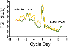 Figure 1. The classic representation of
the ovulatory cycle normalizes hormonal values (in this example, FSH values)
to the day of ovulation. Note that data points corresponding to follicular
phase day 3 (shown in red) for the two subjects pictured are not aligned
in this representation due to differences in their cycle lengths.
Figure 1. The classic representation of
the ovulatory cycle normalizes hormonal values (in this example, FSH values)
to the day of ovulation. Note that data points corresponding to follicular
phase day 3 (shown in red) for the two subjects pictured are not aligned
in this representation due to differences in their cycle lengths.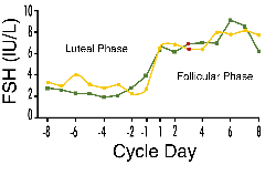 Figure 2. Plot of a portion of the FSH
trajectories for the same two women pictured in Figure 1, normalized to
the luteal-follicular transition. Follicular phase day 3 data points (red)
are aligned in this representation.
Figure 2. Plot of a portion of the FSH
trajectories for the same two women pictured in Figure 1, normalized to
the luteal-follicular transition. Follicular phase day 3 data points (red)
are aligned in this representation.